“Unknown” bacteria (week 2)
My second week in S-STEM was intriguing. I started to get used to the scholars, I began to locate lab equipment’s by myself, and I also stopped getting lost in the laboratories, which is a thumb up for me. This week was all about finding out my “Unknown” bacteria. After being provided my “Unknown” bacteria, figuring out what type (gram positive or gram negative), shape (Cocci, Bacilli), and the name of bacteria was my job to find out. There are multiple steps in finding out your “Unknown” bacteria. Below are the steps I used to demonstrate mine.
1. Streak Plate Method
- Streak plate method is used to isolate colony of bacteria
Materials needed
- “Unknown” bacteria
- Sterilized loop
- TSA plate (Agar plate)
- Parafilm’s
· Methods and Procedures
 |
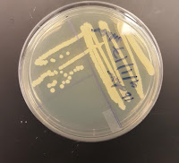
| Figure 1a Colonies of bacteria from 'Streak plate method |
- Using sterilized loops, transfer (dip) your “Unknown” bacteria from a culture (broth) to the TSA plate slightly without pressing in a zigzag manner.
- After your first streaking, sterilize loop and let it cool.
- I repeated the same process (without dipping back again) by rotating (1/4 turn) my TSA plate crossing over streaks from the first streak, and creating another zigzag.
- When done streaking, the TSA plate should be placed in an incubator (37°C) for about twenty-four hours. The bacteria’s will grow, and be in colonies after the streaking method (Figure 1a).
2. Gram Staining Method
- After streaking, it’s now time for gram staining. Cell morphology (the physical feature of cells) are studied by applying multiple stains to cells. Stains are intended to improve physical features of anatomical cell parts. Gram staining differentiates the bacteria between Gram positive (G+) and Gram negative(G-) cell walls.
Materials Needed
- Micro slides
- Sterilized loops
- TSA plate containing ‘Streaked’ bacteria
- Stains (Crystal violet, Grams iodine (IKI), decolorizer, and Safranin)
Methods and procedures
- On a micro slide, add a small drop of water at the center of the slide.
- Using a sterilized loop, transfer a small colony of bacteria to the micro slide and stir/mix gently until you can see a cloud forming.
- When cloud forms, air dry the micro slides for the water to dry off.
 |
| Figure 1b |
- When the water dry’s off, for assurance purposes, apply heat to the micro slide (warm not hot) so the bacteria’s outer part (Glycocalyx) will stick to the micro slide. When applying heat, the bacteria should not face the heat (incinerator).
- The staining process is now under way (Figure 1b).
· Results
 |
| Figure 2a Gram-positive bacteria (1000x) |
- My results came out strong. Under the microscope (1000x magnification), I found out that my “Unknown” bacteria were a ‘Gram positive' (Violet stain after Safranin) 'cocci' (round) bacteria (figure 2a). After finding out my “Unknown” bacteria’s cell wall type and shape, the next process is to conduct different tests (figure 2b) to determine exactly what it is (Genus, Species, and Family).
 |
| Figure 2b Tests to conduct |
· Catalase Test
The first test conducted was a catalase test.
Materials needed
- Micro slide
- Hydrogen peroxide
- Sterilized loop
- Colony of Gram positive bacteria
- A catalase test is made by adding hydrogen peroxide (H2O2) on a micro slide containing a colony of Gram positive bacteria. My bacteria tested positive, hence, created bubbles (figure 2c), which led to my next test.
· Glucose Fermentation
The second test conducted was to see if ‘Glucose’ fermented on my bacteria.
Materials needed
- Glucose (figure 3a)
- Sterilized loop
Methods and Procedure
- Glucose fermentation test is used to see whether your colony of bacteria will change the color of the glucose. A colony of bacteria was added to the glucose container (figure 3a). After twenty-four hours in an incubator, the glucose container changed color like my “Unknown” bacteria did (figure 3b), meaning testing positive for fermenting glucose. You can also notice a little bubble (CO2) on the inverted Durham, which can be concluded as my Gram-positive bacteria being acidic. Once it tested positive for fermentation, it brought me to my final test.
 |
| Figure 3a Glucose before colony of bacteria |
 |
| Figure 3b Glucose after colony of bacteria |
· MSA test (mannitol salt agar)
The third and last test was an MSA test.
Materials Needed
- MSA plate
- Sterilized loop
- Gram-positive bacteria
Methods and procedure
- Taking my ‘Gram-positive’ colony of bacteria, a streak method was used on the MSA plate. The MSA plate was originally pink (figure 4a), but since my bacteria was ‘Gram-positive,’ after twenty-four hours of incubation, it changed the color of the MSA plate to yellow (figure 4b).
- My Gram-positive bacteria tested positive for fermentation making it a Staphylococcus aureus.
 |
| Figure 4a MSA plate before Gram-positive bacteria |
 |
| Figure 4b MSA plate after Gram-positive bacteria |

Comments
Post a Comment